
At-home laboratory test
GI MAP
A stool test combining microbial genetic targets with intestinal-health measurements. Collect your sample at home with the supplied materials and return it to the laboratory. Your written Health Map is included.
Your digestion has been unsettled for a while, or you want to look more closely at an earlier stool result. GI MAP looks for bacteria, parasites and yeast in stool and measures markers of digestion and intestinal inflammation, including the digestive enzyme elastase and the inflammation marker calprotectin. You receive the laboratory report and a written Health Map that explains the results alongside your questionnaire answers and any other results you share. Finding an organism does not establish that it is causing your symptoms.
Price includes delivery, prepaid sample return to the laboratory, laboratory analysis and your written Health Map report.
Pay by card or TWINT
- Sample
- Stool
- Laboratory
- Diagnostic Solutions Laboratory · qPCR
- Laboratory turnaround
- About 21 days after laboratory receipt
Inside your sample report
Read the report before you order.
Follow the actual sample pages, with synthetic values. Your laboratory results come with a personalised written Health Map.
Open the full sample report (PDF) ↗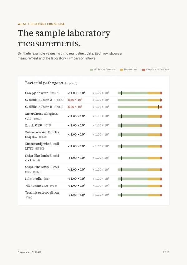
Section 01
Microbial targets
The first results identify the named bacterial targets. Molecular stool testing looks for genetic material. The toxin-labelled entries refer to the target genes listed in the panel. [1]
Section 02
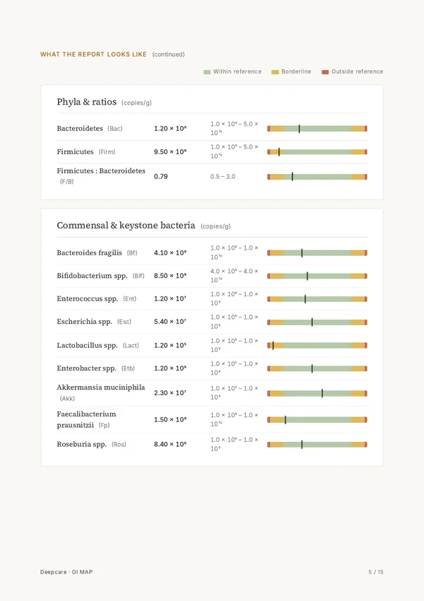
Bacterial groups and individual organisms.
Bacteroidetes and Firmicutes appear as broad groups. Bifidobacterium, Akkermansia and the other named organisms have separate results. The ratio divides the measured Firmicutes quantity by Bacteroidetes.
Section 03
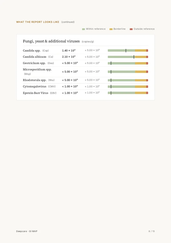
Fungi and the additional viral targets.
Candida species and the other fungal entries sit alongside the additional viral targets. Further pages list protozoa and worms separately, preserving the laboratory's grouping.
Section 04
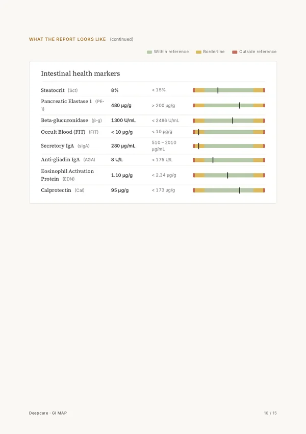
The intestinal-health measurements.
Calprotectin is a protein from inflammatory cells and is used as a stool marker of intestinal inflammation. Pancreatic elastase adds a measurement related to pancreatic digestive-enzyme output. The same page lists the other intestinal markers separately. [2][3]

Section 05
Your written explanation of the results.
Your Health Map is prepared from the results, questionnaire answers and email exchanges. It explains the measurements in the context of the questions you shared with Deepcare.
The complete panel
What the laboratory measures.
Open a group to read each measurement. The groups follow the laboratory panel.
Bacterial pathogens12
- Campylobacter
- Campylobacter commonly reaches people through undercooked poultry and can cause diarrhoea, fever and abdominal cramps. [4]
- C. difficile Toxin A
- Toxin A can damage the intestinal lining by disrupting proteins that organise a cell's internal skeleton. [5]
- C. difficile Toxin B
- Toxin B weakens the bowel lining by disabling proteins that help maintain the shape and internal structure of intestinal cells. [5]
- Enterohemorrhagic E. coli
- These E. coli strains produce Shiga toxins and can cause bloody diarrhoea. [6]
- E. coli O157
- O157:H7 is a well-known Shiga-toxin-producing E. coli serotype involved in foodborne outbreaks. [6]
- Enteroinvasive E. coli / Shigella
- Enteroinvasive E. coli and Shigella can invade the bowel lining and cause inflammatory diarrhoea. [6]
- Enterotoxigenic E. coli LT/ST
- Enterotoxigenic E. coli produces heat-labile or heat-stable enterotoxins and is a common cause of travellers' diarrhoea. [6]
- Shiga-like Toxin E. coli stx1
- The stx1 gene encodes Shiga toxin 1, which can stop protein production inside human cells. [7]
- Shiga-like Toxin E. coli stx2
- Shiga toxin 2 is particularly associated with haemolytic uraemic syndrome, a complication involving damage to small blood vessels and the kidneys. [7]
- Salmonella
- Salmonella can spread through contaminated food or contact with animals, including reptiles and backyard poultry. [8]
- Vibrio cholerae
- Toxin-producing Vibrio cholerae spreads through contaminated food or water and can cause profuse watery diarrhoea with rapid dehydration. [9]
- Yersinia enterocolitica
- Yersinia enterocolitica infection is often linked to raw or undercooked pork and can cause abdominal pain that resembles appendicitis. [10]
Parasitic & viral pathogens5
- Cryptosporidium
- Cryptosporidium infects cells at the surface of the intestine and can spread through water contaminated with its hardy oocysts. [11]
- Entamoeba histolytica
- Entamoeba histolytica is an amoeba that can invade the colon wall and sometimes travel to the liver. [12]
- Giardia
- Giardia attaches to the small-intestinal lining and can interfere with nutrient absorption after transmission through contaminated water or food. [13]
- Adenovirus 40/41
- Adenovirus types 40 and 41 are intestinal adenoviruses known for causing gastroenteritis, particularly in children. [14]
- Norovirus GI/II
- Noroviruses spread readily between people and through contaminated food or surfaces, often causing sudden vomiting and diarrhoea. [15]
H. pylori & virulence factors9
- Helicobacter pylori
- Helicobacter pylori colonises the stomach lining and can cause persistent gastritis and peptic ulcers. [16]
- Virulence factor babA
- BabA is an attachment protein that helps H. pylori bind to blood-group-related sugars on stomach cells. [16]
- Virulence factor cagA
- H. pylori can inject CagA into stomach cells, where it alters signalling and cell structure. [16]
- Virulence factor dupA
- The dupA gene is related to a component of the bacterial type IV secretion machinery, which transfers material between cells. [16]
- Virulence factor iceA
- The iceA1 variant takes its name from its increased expression when H. pylori contacts stomach epithelial cells. [16]
- Virulence factor oipA
- OipA is an outer-membrane protein involved in H. pylori attachment and inflammatory signalling in stomach cells. [16]
- Virulence factor vacA
- VacA is a toxin that can create large fluid-filled compartments inside stomach cells. [16]
- Virulence factor virB
- VirB proteins form parts of the type IV secretion apparatus used by H. pylori to interact with host cells. [17]
- Virulence factor virD
- VirD4 is a component of the H. pylori secretion machinery required for transferring CagA into host cells. [17]
Phyla & ratios3
- Bacteroidetes
- Bacteroidetes include many gut bacteria that break down complex carbohydrates reaching the large intestine. [18]
- Firmicutes
- Firmicutes are a major bacterial group in the human gut and include organisms that produce short-chain fatty acids during fermentation. [18]
- Firmicutes : Bacteroidetes ratio
- This calculation compares the measured quantities of two broad bacterial groups, Firmicutes and Bacteroidetes.
Commensal & keystone bacteria9
- Bacteroides fragilis
- Bacteroides fragilis is an anaerobic colon resident that can cause abscesses if it escapes into tissues outside the bowel. [19]
- Bifidobacterium spp.
- Some Bifidobacterium species can use the complex sugars in human milk, helping explain their abundance in breastfed infants. [18]
- Enterococcus spp.
- Enterococci normally inhabit the intestine but can cause infections when they reach the bloodstream or urinary tract. [20]
- Escherichia spp.
- Escherichia coli is a common intestinal resident, with distinct strains responsible for urinary and intestinal infections. [21]
- Lactobacillus spp.
- Lactobacilli ferment sugars into lactic acid and occur in fermented foods as well as human mucosal communities. [22]
- Enterobacter spp.
- Enterobacter species can inhabit the digestive tract and are also recognised causes of infections associated with hospital care. [21]
- Akkermansia muciniphila
- Akkermansia muciniphila specialises in using mucin, the main protein component of the mucus covering the gut lining. [18]
- Faecalibacterium prausnitzii
- Faecalibacterium prausnitzii produces butyrate, a short-chain fatty acid used as fuel by cells lining the colon. [18]
- Roseburia spp.
- Roseburia species ferment dietary carbohydrates to butyrate and are studied for their contribution to colonic metabolism. [23]
Opportunistic & overgrowth bacteria9
- Bacillus spp.
- Bacillus species form resistant spores, and some foodborne species can produce toxins that cause vomiting or diarrhoea. [24]
- Enterococcus faecalis
- Enterococcus faecalis can move from its normal intestinal habitat to cause urinary tract infections or infection of the heart valves. [20]
- Enterococcus faecium
- Enterococcus faecium is a gut commensal whose hospital-adapted lineages are prominent in research on antimicrobial resistance. [20]
- Morganella spp.
- Morganella morganii can inhabit the intestine and occasionally causes opportunistic urinary, wound or bloodstream infections. [25]
- Pseudomonas spp.
- Pseudomonas species are widespread in water and soil and can temporarily colonise human skin or the digestive tract. [26]
- Pseudomonas aeruginosa
- Pseudomonas aeruginosa is particularly associated with opportunistic infections in people with burns, cystic fibrosis or weakened immune defences. [26]
- Staphylococcus spp.
- Many staphylococci normally live on human skin, where disruption of the skin barrier can give them access to deeper tissues. [27]
- Staphylococcus aureus
- Staphylococcus aureus commonly colonises the nose and can produce enterotoxins responsible for food poisoning. [27]
- Streptococcus spp.
- Streptococci include normal inhabitants of the mouth and throat as well as species that cause respiratory or invasive infections. [28]
Methanogens & sulphate reducers2
Additional bacteria9
- Citrobacter spp.
- Citrobacter belongs to the coliform bacteria found in the intestine and can cause opportunistic infections outside its usual habitat. [21]
- Citrobacter freundii
- Citrobacter freundii is one of the intestinal coliform species implicated in urinary tract and bloodstream infections. [21]
- Klebsiella spp.
- Klebsiella species have a protective capsule and can colonise the intestine before causing infections at other body sites. [21]
- Klebsiella pneumoniae
- Klebsiella pneumoniae is an encapsulated organism associated with pneumonia as well as urinary and bloodstream infections. [21]
- M. avium subsp. paratuberculosis
- This mycobacterium causes Johne's disease, a chronic intestinal infection of ruminants such as cattle. [30]
- Proteus spp.
- Proteus species can live in the gut and are recognised opportunistic causes of urinary tract infections. [21]
- Proteus mirabilis
- Proteus mirabilis produces urease, an enzyme that can raise urine pH and contribute to infection-related urinary stones. [21]
- Fusobacterium spp.
- Fusobacteria are anaerobic organisms found in the mouth and intestine and can take part in infections involving several bacterial species. [19]
- Prevotella spp.
- Prevotella species inhabit mucosal sites including the mouth and can participate in anaerobic infections after tissue barriers are breached. [19]
Fungi, yeast & additional viruses7
- Candida spp.
- Candida yeasts can inhabit the digestive tract as commensals and cause opportunistic infections when local conditions or immune defences change. [31]
- Candida albicans
- Candida albicans can switch between yeast and filamentous forms, a feature involved in its interactions with human tissues. [31]
- Geotrichum spp.
- Geotrichum is a yeast-like fungus found in the environment and occasionally as part of the human intestinal flora. [32]
- Microsporidium spp.
- Microsporidia are spore-forming intracellular parasites that can cause persistent diarrhoea, particularly in people with weakened immunity. [33]
- Rhodotorula spp.
- Rhodotorula species are pigmented environmental yeasts known for occasional catheter-associated bloodstream infections. [34]
- Cytomegalovirus
- Cytomegalovirus remains in the body after infection and can reactivate when immune defences are weakened. [35]
- Epstein-Barr Virus
- Epstein-Barr virus commonly spreads through saliva and is the main cause of infectious mononucleosis. [36]
Protozoa7
- Blastocystis hominis
- Blastocystis is a single-celled intestinal organism found in people both with and without digestive symptoms. [37]
- Chilomastix mesnili
- Chilomastix mesnili lives in the caecum or colon and spreads when its cysts are swallowed in contaminated food or water. [38]
- Cyclospora spp.
- Cyclospora cayetanensis infects the small intestine and has caused outbreaks linked to fresh produce. [39]
- Dientamoeba fragilis
- Dientamoeba fragilis is a single-celled parasite of the large intestine that occurs in both symptomatic and symptom-free carriers. [40]
- Endolimax nana
- Endolimax nana is a small intestinal amoeba acquired by swallowing mature cysts from faecally contaminated material. [41]
- Entamoeba coli
- Entamoeba coli is a colon-dwelling amoeba whose mature cysts typically contain eight nuclei. [41]
- Pentatrichomonas hominis
- Pentatrichomonas hominis is a flagellated organism that lives in the large intestine and has no known cyst stage. [42]
Worms (helminths)5
- Ancylostoma duodenale
- Ancylostoma duodenale is a hookworm that attaches to the small intestine and feeds on blood, contributing to iron loss. [43]
- Ascaris lumbricoides
- Ascaris larvae migrate through the lungs before returning to the small intestine, where the adult worms live. [44]
- Necator americanus
- Necator americanus larvae usually enter through bare skin exposed to contaminated soil before developing into blood-feeding intestinal worms. [43]
- Trichuris trichiura
- Trichuris trichiura is the whipworm, which embeds its thin front end in the lining of the caecum and colon. [45]
- Taenia spp.
- Humans acquire intestinal Taenia tapeworms from raw or undercooked beef or pork containing larval cysts. [46]
Intestinal health markers8
- Steatocrit
- Steatocrit is a semiquantitative measurement of fat in stool, expressed as the fat fraction of the processed sample. [47]
- Pancreatic Elastase 1
- Pancreatic elastase reaches the stool after being released with pancreatic digestive enzymes, so its concentration is used to assess exocrine pancreatic function. [3]
- Beta-glucuronidase
- Gut bacterial beta-glucuronidases are enzymes that remove a sugar-like group the body has attached to substances for excretion, sometimes allowing those substances to be absorbed again. [48]
- Occult Blood (FIT)
- FIT uses antibodies to measure human haemoglobin in stool, including blood present in amounts too small to see. [49]
- Secretory IgA
- Secretory IgA is an antibody that binds microbes and their components inside the intestine, helping keep them away from the gut lining. [50]
- Anti-gliadin IgA
- This stool measurement counts IgA antibodies that bind gliadin, a protein fraction of gluten.
- Eosinophil Activation Protein (EDN/EPX)
- Eosinophil-derived neurotoxin is an enzyme that breaks down RNA, the molecule cells use to make proteins, and is released when immune cells called eosinophils are activated. [51]
- Calprotectin
- Calprotectin is a protein abundant in immune cells called neutrophils, so increased amounts in stool reflect intestinal inflammation involving these cells. [2]
Scientific references
- 2017 Infectious Diseases Society of America Clinical Practice Guidelines for the Diagnosis and Management of Infectious Diarrhea. Shane AL, Mody RK, Crump JA, Tarr PI, Steiner TS, Kotloff K, Langley JM, Wanke C, Warren CA, Cheng AC, Cantey J, Pickering LK., 2017. DOI: 10.1093/cid/cix669. PMID: 29053792.
- Role of fecal calprotectin as a biomarker of intestinal inflammation in inflammatory bowel disease. Konikoff MR, Denson LA., 2006. DOI: 10.1097/00054725-200606000-00013. PMID: 16775498.
- AGA Clinical Practice Update on the Epidemiology, Evaluation, and Management of Exocrine Pancreatic Insufficiency: Expert Review. Whitcomb DC, Buchner AM, Forsmark CE., 2023. DOI: 10.1053/j.gastro.2023.07.007. PMID: 37737818.
- About Campylobacter infection. Centers for Disease Control and Prevention, accessed 2026.
- Clostridioides difficile toxins: mechanisms of action and antitoxin therapeutics. Kordus SL, Thomas AK, Lacy DB., 2022. DOI: 10.1038/s41579-021-00660-2. PMID: 34837014.
- FAQ: E. Coli: Good, Bad, & Deadly. American Academy of Microbiology, 2011.
- Shiga Toxin (Stx) Classification, Structure, and Function. Melton-Celsa AR., 2014. DOI: 10.1128/microbiolspec.EHEC-0024-2013. PMID: 25530917.
- About Salmonella Infection. Centers for Disease Control and Prevention, accessed 2026.
- Cholera. World Health Organization, accessed 2026.
- About Yersinia Infection. Centers for Disease Control and Prevention, accessed 2026.
- DPDx: Cryptosporidiosis. Centers for Disease Control and Prevention, accessed 2026.
- DPDx: Amebiasis. Centers for Disease Control and Prevention, accessed 2026.
- DPDx: Giardiasis. Centers for Disease Control and Prevention, accessed 2026.
- Guidelines for Outbreaks: Adenovirus. Centers for Disease Control and Prevention, accessed 2026.
- About Norovirus. Centers for Disease Control and Prevention, accessed 2026.
- Virulence Factors of Helicobacter pylori: A Review. Roesler BM, Rabelo-Gonçalves EM, Zeitune JM., 2014. DOI: 10.4137/CGast.S13760. PMID: 24833944.
- Architecture of the Helicobacter pylori Cag-type IV secretion system. Terradot L, Waksman G., 2011. DOI: 10.1111/j.1742-4658.2011.08037.x. PMID: 21352491.
- Introduction to the human gut microbiota. Thursby E, Juge N., 2017. DOI: 10.1042/BCJ20160510. PMID: 28512250.
- Anaerobic Gram-Negative Bacilli. Hentges DJ, 1996.
- Enterococci and Their Interactions with the Intestinal Microbiome. Dubin K, Pamer EG., 2017. DOI: 10.1128/microbiolspec.BAD-0014-2016. PMID: 29125098.
- Escherichia, Klebsiella, Enterobacter, Serratia, Citrobacter, and Proteus. Guentzel MN, 1996.
- Lactobacilli biology, applications and host interactions. Mejía-Caballero A, Marco ML, 2025. DOI: 10.1038/s41579-025-01205-7.
- Roseburia spp.: a marker of health?. Tamanai-Shacoori Z, Smida I, Bousarghin L, Loreal O, Meuric V, Fong SB, Bonnaure-Mallet M, Jolivet-Gougeon A., 2017. DOI: 10.2217/fmb-2016-0130. PMID: 28139139.
- Bacillus. Turnbull PCB, 1996.
- Morganella morganii, a non-negligent opportunistic pathogen. Liu H, Zhu J, Hu Q, Rao X., 2016. DOI: 10.1016/j.ijid.2016.07.006. PMID: 27421818.
- Pseudomonas. Iglewski BH, 1996.
- Staphylococcus. Foster T, 1996.
- Streptococcus. Patterson MJ, 1996.
- Hydrogen cross-feeders of the human gastrointestinal tract. Smith NW, Shorten PR, Altermann EH, Roy NC, McNabb WC., 2019. DOI: 10.1080/19490976.2018.1546522. PMID: 30563420.
- Mycobacterium avium subsp. paratuberculosis in Veterinary Medicine. Harris NB, Barletta RG., 2001. DOI: 10.1128/CMR.14.3.489-512.2001. PMID: 11432810.
- Mixed Mycotic Infections. Soll DR, 2002.
- Fungi in the healthy human gastrointestinal tract. Hallen-Adams HE, Suhr MJ., 2017. DOI: 10.1080/21505594.2016.1247140. PMID: 27736307.
- DPDx: Microsporidiosis. Centers for Disease Control and Prevention, accessed 2026.
- ESCMID and ECMM joint clinical guidelines for the diagnosis and management of rare invasive yeast infections. Arendrup MC et al., 2014. DOI: 10.1111/1469-0691.12360.
- About Cytomegalovirus. Centers for Disease Control and Prevention, accessed 2026.
- About Epstein-Barr Virus. Centers for Disease Control and Prevention, accessed 2026.
- DPDx: Blastocystis. Centers for Disease Control and Prevention, accessed 2026.
- DPDx: Chilomastix mesnili. Centers for Disease Control and Prevention, accessed 2026.
- DPDx: Cyclosporiasis. Centers for Disease Control and Prevention, accessed 2026.
- DPDx: Dientamoeba fragilis. Centers for Disease Control and Prevention, accessed 2026.
- DPDx: Intestinal Amebae. Centers for Disease Control and Prevention, accessed 2026.
- DPDx: Non-pathogenic Flagellates. Centers for Disease Control and Prevention, accessed 2026.
- DPDx: Intestinal Hookworm. Centers for Disease Control and Prevention, accessed 2026.
- DPDx: Ascariasis. Centers for Disease Control and Prevention, accessed 2026.
- DPDx: Trichuriasis. Centers for Disease Control and Prevention, accessed 2026.
- DPDx: Taeniasis. Centers for Disease Control and Prevention, accessed 2026.
- Steatocrit: a reliable semiquantitative method for detection of steatorrhea. Sugai E, Srur G, Vazquez H, Benito F, Mauriño E, Boerr LA, Bai JC., 1994. DOI: 10.1097/00004836-199410000-00007. PMID: 7806830.
- The role of gut microbial β-glucuronidase in drug disposition and development. Gao S, Sun R, Singh R, Yu So S, Chan CTY, Savidge T, Hu M., 2022. DOI: 10.1016/j.drudis.2022.07.001. PMID: 35820618.
- Recommendations on fecal immunochemical testing to screen for colorectal neoplasia: a consensus statement by the US Multi-Society Task Force on colorectal cancer. Robertson DJ, Lee JK, Boland CR, Dominitz JA, Giardiello FM, Johnson DA, Kaltenbach T, Lieberman D, Levin TR, Rex DK., 2017. DOI: 10.1016/j.gie.2016.09.025. PMID: 27769516.
- Multi-faceted functions of secretory IgA at mucosal surfaces. Corthésy B., 2013. DOI: 10.3389/fimmu.2013.00185. PMID: 23874333.
- Eosinophil granule proteins: form and function. Acharya KR, Ackerman SJ., 2014. DOI: 10.1074/jbc.R113.546218. PMID: 24802755.
Add a SIBO breath test
CHF 199, only with GI MAP. One breath test per GI MAP.
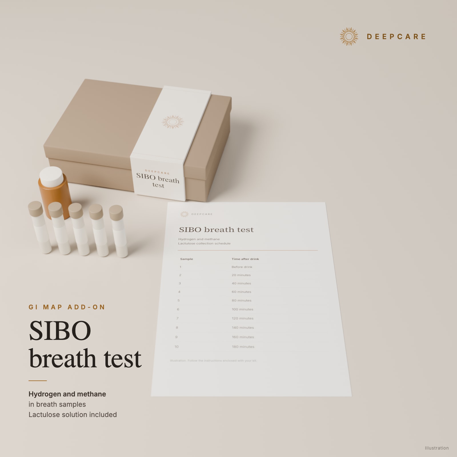
If you have persistent bloating, gas, abdominal pain, diarrhoea or constipation, discuss with your doctor whether breath testing fits your symptoms and history. SIBO means an excess of bacteria in the small intestine. This home test measures hydrogen and methane in your breath after a lactulose drink.
Add GI MAP to your basket, then choose the breath test there. ↗
Who is breath testing for?
The 2020 American College of Gastroenterology guideline suggests breath testing for people with irritable bowel syndrome, symptoms with a suspected intestinal movement disorder, or symptoms after surgery involving the digestive tract. These are conditional recommendations based on very low-quality evidence. It also suggests methane testing for people with constipation and symptoms, and advises against testing people without symptoms solely because they take proton-pump inhibitors, medicines that reduce stomach acid. [1]
The 2017 North American consensus supports testing for unexplained bloating and gas, and methane measurement with constipation. Guidance differs: a 2024 update endorsed by the European and American neurogastroenterology societies argues against using breath tests to establish SIBO in irritable bowel syndrome. Lactulose can reach the colon quickly and produce a false positive. [2] [3]
How collection works at home
Allow 3 hours for collection. Take a baseline breath sample, then drink the supplied 10 g lactulose mixed with 230 mL water within 5 minutes. Collect another sample every 20 minutes until 180 minutes, for 10 tubes in total. Do not drink water for the first hour after the drink. Keep the sealed tubes at room temperature and dispatch them within 2 days, following the enclosed instructions. [4]
Preparation and restrictions
Follow the instructions enclosed with your kit. Never stop a prescribed medicine without your doctor's agreement. Discuss conflicting medicines or special diets before ordering.
- Wait at least 4 weeks after antibiotics, diarrhoea, colonoscopy or a barium examination/enema; 2 to 4 weeks after antifungals, bismuth subsalicylate (Pepto-Bismol) or herbal antimicrobials.
- For 7 days beforehand, avoid laxatives, stool softeners, bulking agents and antacids containing aluminium or magnesium hydroxide.
- In the preceding 24 hours, stop probiotics. Before the fast, eat only baked or grilled chicken, fish or turkey with salt and pepper, white bread, plain white rice, eggs or clear chicken/beef broth without vegetables. Drinks: plain water, coffee or tea without sugar, sweeteners, milk or cream.
- For 12 hours before and throughout the test, fast: water only, with the first-hour exception above. Avoid non-essential medicines and supplements unless advised otherwise, chewing gum, sweets and mouthwash.
- For 1 hour before and during collection: no smoking or second-hand smoke, sleeping, vigorous exercise or toothpaste.
The instructions advise against this test after an allergic reaction to lactulose or with a medically required galactose- or lactulose-restricted diet, and they restrict it for some people with diabetes. If you have diabetes, ask your doctor before ordering. If you cannot follow the preparation, discuss another test with your doctor. [4]
What you receive
The collection kit includes the lactulose solution, 10 tubes, labels and a breath-sampling device. Laboratory analysis, delivery and prepaid return are included. You receive the breath-test results and one written Health Map that considers them together with your GI MAP results, questionnaire answers and other results you share. There is no separate Health Map for the breath test.
A positive result is a finding to take to a doctor, who decides whether any treatment is needed. Breath testing can give false positive or false negative results. Methane comes from methane-producing microorganisms that can live in both the small and large intestine; a methane-positive result does not locate them or prove bacterial overgrowth in the small intestine. [1] [5]
Sources for the breath test
- ACG Clinical Guideline: Small Intestinal Bacterial Overgrowth. Pimentel et al., 2020.
- Hydrogen and Methane-Based Breath Testing in Gastrointestinal Disorders: The North American Consensus. Rezaie et al., 2017.
- Critical appraisal of the SIBO hypothesis and breath testing. Kashyap et al., 2024.
- Collection instructions enclosed with the kit (English).
- European guideline on hydrogen and methane breath tests. Hammer et al., 2022.
Included in the price
Your kit and your written Health Map.
Delivery to Switzerland, Liechtenstein and every EU country in 2 to 3 working days. Sample return is included.
The kit arrives in the laboratory's packaging. Product pictures are illustrations.
CHF 399
Price includes delivery, prepaid sample return to the laboratory, laboratory analysis and your written Health Map report.
Pay by card or TWINT
Add a SIBO breath test · CHF 199 ↗
With this kit, add OAT Usual price: CHF 369 Combo price: CHF 319 or DUTCH Complete Usual price: CHF 399 Combo price: CHF 349.
Your written Health Map report
The kit price includes a systematic written evaluation for each person tested. We consider all of that person's kit results together with their questionnaire answers and any other results they share. After payment, your first personalised questionnaire arrives by email. Your answers, laboratory results and email exchanges form the basis of the report. Follow-up questions arrive by email or a further questionnaire when needed. The final report is written in your language, English, French or German, within 3 working days of your answers to the last questionnaire.
Read about the Health Map ↗Questions about this kit
Does this kit include zonulin?
This kit is the GI MAP panel without the zonulin add-on. The full list below describes the included panel.
How do I return the sample?
Use the collection container and return materials supplied in the kit. Follow its packing and shipping instructions, including any cooling instructions. Sample return is included in the price.